And so it begins...

I love science fiction. As both a Star Wars fan and a scientist, I often find myself daydreaming of what the future holds for both technology and mankind. As we continue to progress as humans, we will inevitably hit numerous ethical speed bumps (or speed mountains) along the way to becoming the most advanced species in the universe.  Want to get to Mars? Live forever? Cure all human disease? All of these things sound great, but these advancements are always tied to some pretty hard choices.

Figure 1: Sometimes the choice won't be this easy! The answer is cake, by the way.

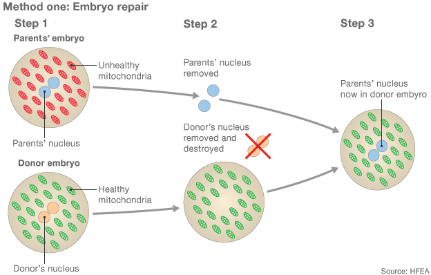
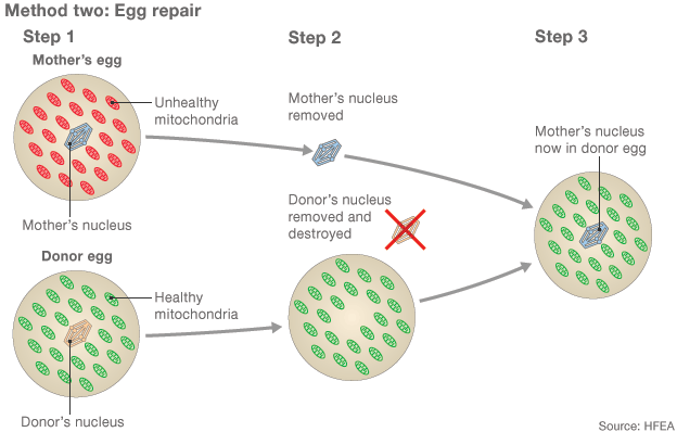
A few months ago, I blogged about how genetic engineering has the potential to help cure a variety of human diseases. I also mentioned how thought leaders in the genetic engineering space proposed that a moratorium be held on any attempts to use such technology to edit a human embryo. Well it seems like that those requests were ignored, as a study was recently published in Protein and Cell attempting to correct the gene that causes beta thalassemia in embryos. This disease results in misshapen hemoglobin, the component of your blood that carries oxygen. Misshapen hemoglobin leads to anemia, so you can see why someone would want to cure it! In the above paper, 86 embryos were used in an attempt to correct the mutation. They used CRISPR/Cas9, a very clever system discovered in 2012 that has the ability to hone in on specific regions of DNA, cut out the gene in question, and replace it with a different one. Only a small number correctly replaced the mutated gene with a normal copy, and the process also generated mutations in other unintended parts of the genome.

Let me be very clear here. The researchers (at least, to my knowledge) never intended to implant these embryos into an actual womb. As controversial as this study was, it actually provides us with important confirmation of what we already suspected - we simply need to learn more about how gene editing technologies like CRISPR/Cas9 work before we can hope to use them in an actual embryo. The success rate for this paper was pretty small. The success rate if we want to start using this routinely? 100%.

This technology has the potential to rid the human race of a whole host of genetic disorders, but the fact that we are still extraordinarily naive about how exactly such technologies will behave in an actual embryo cannot be understated. The majority of scientific institutions throughout the world seem to agree with me, with over 170 companies, patient groups, and research institutions stating that a moratorium is necessary. Nature and Science, two of the world's most influential journals, rejected the paper due to its ethical dubiousness.

Just as choosing whether or not to pursue such research will always remain a difficult choice, it is inevitable that this sort of research will continue to be attempted. It doesn't matter if 1000 research centers say that it is dangerous - it only takes on lab. The argument can certainly made that unless we try these techniques in actual humans, we'll never truly know if they work or not. There is only so much that you can do in a laboratory before one needs to make the scientific leap. This leap has been taken many times throughout the history of science, and it's always fraught with the unknown.

Take, for example, Edward Jenner. His name may not be familiar to you, but his invention of the vaccine has saved millions of people around the world. How did he discover this? He inoculated the 8 year old son of his gardener with pus from a person with cowpox, then challenged him with the virus. This worked beautifully, but it certainly seems a little ethically questionable, don't you think?

Figure 2: Edward Jenner advising a farmer to vaccinate his family

Of course, we know better now. We have better informed science, and we have better models upon which to test our ideas. So when will we see the successful correction of a mutation in a human embryo? Only time will tell, but I can guarantee that it'll happen sooner than you think!

Transduction, s'il vous plait?

As most of you are aware, I work for the National Center for Advancing Translational Science (NCATS) here in Rockville, MD. We often talk about translational research at the office - how it works, how we can improve on it, and how we can use it to make people healthier. But what is translational research? The vast majority of biomedical research in the United States is known as basic research. This research seeks to understand the fundamental mechanisms that govern life - how genes are regulated, what proteins influence biochemical pathways in the body, and so on and so forth. I think that this research needs relabeling, as basic seems to imply a lack of complexity (at least, that's what it sounds like to me!). The word fundamental would probably be more apt here, as this research is absolutely critical to applied research. Applie research uses the information gathered in basic research to create new therapies for humans, such as new medical devices or new drugs. The process by which knowledge is transformed from basic to applied research is known as translation, which is the challenging topic that my institute focuses on.

This is certainly easier said than done. As you can imagine, a vast amount of basic research is performed around the world every year, yet the rate at which we develop new therapies is much lower. 200 billion USD was spent on research and development last year ($90 billion in the US) alone in the life sciences, while thousands of scientists work around the clock to figure out new ways to tackle problems in human health. Do you know how many drugs were approved by the FDA last year? 41. Forty. One. This is actually much higher than the 21 per year that the FDA averaged over the past decade.

Okay, let me be a bit transparent here. That figure is of novel drugs approved, which means that the drugs were completely brand-spankin' new. This would be in contrast to repurposed drugs, which are drugs that have already been developed by can now be used for another disease. But even if that number of drugs were in the hundreds, it is still abysmally low relative to the amount of money that we put into research. What gives?

Have you ever volunteered for a walk to help fund medical research? Maybe you've donated some money here and there to foundations promising to accelerate research. If you've been in either of those scenarios, I'm sure you've felt frustrated that it seems like the amount of energy and money that we spend trying to cure diseases usually results in very limited results.

In the end, it boils down to one thing: it's really really really really really really x 1000 difficult to move an idea found in the laboratory to the clinic. So difficult in fact, that my institute was founded on the very premise of solving this as a systematic problem. Once we can solve this problem, we will be able to utilize the vast wealth of basic research that scientists around the world have spent their entire careers on. As the Isaac Newton once said, "If I have seen further it is by standing on the shoulders of giants".

Before I let you go, I'm going to spoil the ending a little. Despite the sort of dreary tone this blog post holds, I am infinitely optimistic that we will only continue to improve the way in which we discovery new drugs and develop new medical interventions. Costs will go down, precision will go up, and most importantly, the quality of life for sick patients around the world will soar. But before we can cross the quagmire that is translation, we're going to need some supplies. What makes translation so difficult? Where do we need to improve on? This is what I'll be discussing in the next few blog posts, so stay tuned for more!

Achoo! How drugs saved my life.

I've been over winter since about mid-January, so when March 20th rolled around I was more than happy to greet the new season. Cherry blossoms, warm weather, and warm rain showers. Best season ever, right? WRONG. WRONG. WRONG. WRONG.

I love most of spring as much as the next guy, but when those pollen counts start ticking higher and higher my body begins a heavy revolt. I start to sneeze, my nose runs, my eyes itch, and my mind starts to become overwhelmed. It heavily affects my ability to focus and is an overall huge hindrance to my productivity - not good when you're working in a lab! For today's post, I'm going to focus on something that has kept me from having a meltdown over the past few weeks - antihistamines.

Figure 1. This is why I don't like going outside. Yuck!

When you experience and allergy attack, be it hives or a runny nose, your body is mounting an immune response to a perceived threat. My immune system, for example, hates cats, grass, and trees. When it detects bits and pieces from these things in the form of dander and pollen, it ramps up the production of antibodies against these allergens. These antibodies then bind to cells called mast cells which contain large amounts of histamine. Once released, this histamine causes a variety of the symptoms you normally associate with an allergic response. For example, these antibodies can bind to mucous membranes in your nasal cavity, leading to a histamine release which causes sneezing and nasal congestion.

Antihistamines work by blocking the receptors that recognize histamine, resulting in a reduced immune response. The earliest antihistamines developed, also known as first generation antihistamines, were not very specific to the histamine receptor. This lead to some undesirable side effects, including drowsiness thanks to their ability to cross the blood-brain barrier into the central nervous system. This is why if you've taken one of these medications (i.e. Benadryl, ChlorTrimeton), you may have had to avoid driving! Because of this side effect, some modifications to these first generation antihistamines are used to treat insomnia (i.e. ZzzQuil).

Then came the second generation antihistamines, which were much better at targeting their intended histamine receptors and only crossed the blood-brain barrier to a small extent. These "non-drowsy" medications include Claritin, Allegra, and Zyrtec. Despite being in the same class of antihistamines, all of these different medications have varying chemical structures and potencies. Your results may vary!

Finally, one of the most popular classes of allergy medications in recent years has been the nasally administered corticosteroids, which includes Flonase and Nasacort. These nasal sprays work in a slightly different way than the above medications and aren't technically antihistamines. These corticosteroids bind to glucocorticoid receptors in the nasal passages, which are important in downregulating immune activity (inflammation).

In my experience, Flonase has worked the best for me. Each person is different, however, and may require different regiments of allergy prevention medications to be effective. For more information, go see your doctor! In the meantime, I'm going to go back into the lab, happy and heavily medicated.

The Epigenome

Today, a slew of very important research papers were published in the journal Nature describing a pretty incredible feat of science - the sequencing of the epigenome (well, mostly). Most of you are probably more familiar with the sequencing of the human genome via the Human Genome Project. Led by the National Institutes of Health (NIH), this endeavor mapped all of the genes in the human body and concluded in the early 2000's. The implications of this new knowledge were pretty huge! From a better understanding of the genetic basis of cancer to more personalized drug therapies, scientists gained a very important milestone in the difficult journey of learning how our bodies work.

Today, we gained another milestone through the sequencing of the epigenome, which are the modifications on the genome that affect which genes are expressed in a given cell. Think for a moment of how diverse the cells in your body can be - some can fire electrical signals to send messages throughout the nervous system (neurons) while some are engineered to produce that insulin that controls your blood glucose (beta cells in your pancreas). The genetic material in these cells, no matter where they are, is exactly the same, give or take the occasional mutation here and there. What makes the cells different from one another are what genes are turned on and off, which is partially controlled by your epigenome.

Figure 1: The epigenome controls which genes are "unwound", revealing their information to your cells. ADAPTED FROM ILLUSTRATION BY SIGRID KNEMEYER, PREVIOUSLY ADAPTED BY LAUREN SOLOMAN

Figure 1: The epigenome controls which genes are "unwound", revealing their information to your cells. ADAPTED FROM ILLUSTRATION BY SIGRID KNEMEYER, PREVIOUSLY ADAPTED BY LAUREN SOLOMAN

This $300 million project, again supported by the NIH, was quite challenging to say the least! With the human genome project, scientists only had to worry about one genome. The epigenome, however, differs from cell to cell, making any attempts to sequence it pretty onerous. This makes sense, as the epigenome is essential for defining an individual cell's identity! The efforts mentioned above sequenced about 111 different tissues, with more to come in the future.

So in the end, why does this matter ? Our knowledge of the epigenome will allow us to notice smaller differences between people at the molecular level, which can lead to a better understanding of what causes human disease. For example, scientists may reexamine patients with cancers caused by genetic mutations and ask themselves "what role does the epigenome play in this disease?". With insight from the now-sequenced epigenome, they may discover that certain genes are inappropriately turned "on" in cancer patients, leading to therapies that target those specific genes.

Keep an eye out for more developments in this field - they're going to be pretty monumental!

What is love? (Baby don't hurt me)

"Do you believe in love at first sight, or should I walk by again?" - Unknown When I sit at my local coffee shop to work on planning experiments, about 15-20% (rough estimation) of the patrons around me appear to be on dates. Fresh off the heels of Valentine's Day, a thought passed through my head that I thought was pretty intriguing - what is love?

Figure 1: Wall-E and EVE. This post may not apply to robots.

Figure 1: Wall-E and EVE. This post may not apply to robots.

Emotion is a classic topic in science that has been somewhat difficult to explain over the centuries. As a biologist, my first instinct is to boil down such a complex social phenomenon into its individual physiological components. The difference between happiness, sadness, jealousy, anger, and everything in between is simply the way in which the electrical signals in your brain coordinate the release of neurotransmitters, like dopamine and GABA. Some see this perspective as rather dull, but I think it's really quite beautiful!

When you see someone very attractive, what happens to you? Your heart begins to race, you start to feel very warm, and it seems like everything surrounding the object of your affection fades into the background. What explains this fixation? A fascinating review in the Journal of Sexual Medicine focused on this specific topic. With the use of advanced technologies, we can better visualize the physiological basis behind that warm and fuzzy feeling you get when you see someone you're interested in!

Ortigue et al. differentiate between two types of love in the review I've hyperlinked above. The first is known as passionate love, which is the sort of love you have towards a significant other. The second is compassionate love, which is the love you might feel towards a good friend or a family member. By using a device known as functional magnetic resonance imaging (fMRI), scientists can see how blood flow throughout the brain changes when these types of love are experienced.

In one study, scientists allowed participants to view images of their respective partners for about 17 seconds, while taking a peak into their brain via fMRI. They found that blood flow was significantly increased in areas of the brain associated with reward and euphoria, including areas of the brain that are also associated with euphoria-inducing drugs like cocaine. Activity was also shown in areas involved in memory. Simultaneously, blood flow was decreased in areas of the brain associated with anxiety and fear, such as the amygdala. These are also the areas of the brain that are highly active in humans who experience emotional stress, like a bad break up. A second study found that the reward centers of the brain are also stimulated when participants were shown the name of their significant other. How romantic!

Bartels and Zeki in 2004 performed a similar study, but this time they were seeking to further understand compassionate love. The experimental design was similar to the one above. In this case, the participants were all mothers who were shown pictures of their children. The researchers found significant activity in an area of the brain known as the periaqueductal gray matter (PAG), an area of the brain that is involved in pain suppression during intense emotional experiences, such as childbirth. Activity was also shown in areas of the brain associated with higher cognitive processing.

The same author has also discussed the idea of "love at first sight", which about 58% of Americans believe is a real phenomenon. As it turns out, when you look at someone who you find extremely attractive, 12 different areas of the brain work together to release a whole host of different neurotransmitters within 0.2 seconds of visual contact. Just think about that - within a fifth of a second, your brain is able to tell you "Wow, this person is incredible!" as opposed to the person to their left or right. Whether or not this feeling of intense emotion is actually "love" is a debate that continues to rage on today. What does this scientist think it means? From a biological perspective, it goes something like this:

"Ah, this person looks like they have good genes. Probably would make strong, healthy offspring. Proceed with mating."

In short, your brain is capable of generating very different feelings of love depending on the situation. It's a pretty incredible system, isn't it? Of course, why some people trigger strong feelings of love over others is a lengthy subject and probably too much for the scope of this blog. So when the next time you see your significant other (or that cute barista working the espresso machine), think about how hard your brain is working to say, "Hey buddy, time to make your move!"

What does it mean to have three parents?

I commented on this briefly in a blog post some time ago, but this topic has seen a resurgence in the media lately. This past week, the British House of Commons voted to allow for the creation of a baby made with the genetic material of three people, so called "three-person babies". Not surprisingly, this has some people up in arms over the ethics of such a procedure, as well as its implications for the future of genetic modification. I even saw someone call it "letting the gene out of the bottle". Clever! In true Kitchen Table Science fashion, I'm going to give you a brief overview of what this technology is and what is it meant to correct. Just the facts ma'am, just the facts!

1. The Mitochondria

You may remember from high school that the mitochondria is the powerhouse of the cell, responsible for the majority of energy generation for your body. You may also remember that it, in fact, has its own genetic material independent of the rest of your genome. The reasoning behind this is theoretical and may be discussed in a future post, but know that the DNA present in your mitochondria comprises less than a tenth of a percent of your overall genetic code. Its 37 (versus the remaining 20-25,000 genes in your genome) genes do not influence anything related to appearance, intelligence, or anything along those lines.

2. Mitochondrial disease

As with all genes, your mitochondrial DNA is susceptible to mutations. These mutations can cause a family of disease known as mitochondrial diseases. Symptoms of these diseases vary wildly and include poor growth, muscle weakness, and poor coordination. Since your mitochondria process DNA separately from the rest of the cell, you can theoretically correct for mitochondrial disease by replacing the damaged organelle with a healthy one in the embryo. The healthy mitochondria would continue to be passed down to the cells in the developing embryo, ensuring that all cells in the fully formed human will have good mitochondria. Below is a great graphic from the Human Fertilization and Embryology Authority on how this works:

Figures 1 and 2: Proposed methods for correcting mitochondrial genetic abnormalities

Figures 1 and 2: Proposed methods for correcting mitochondrial genetic abnormalities

So is this safe and/or ethical? Technically speaking, yes, the resulting child will have the genetic material of three different people. Calling the donor of the mitochondria a "parent" though is a pretty big stretch, considering how little of the DNA contributed will actually affect the rest of the body. It is impossible to predict whether or not something will be completely safe when it comes to procedures like this - only time will be able to tell us that.

I'll leave it for you to decide whether or not society should proceed with this technology. My opinion? This presents an exciting new way to correct for some devastating diseases, and am I certainly looking forward to seeing how this technique evolves in the coming months!

A little vaccine update

Last week, I received a booster dose of the VSV-EBOV Ebola vaccine that I've blogged about before, and I'm happy to report that I had no symptoms this go around. This is a pretty stark contrast from when I got my first dose...a 101.6 degreefever and terrible joint pain make for a pretty miserable evening!

Figure 1: A picture of the vaccine, isn't it cute?

Figure 1: A picture of the vaccine, isn't it cute?

Since starting the trial, a few bits of news regarding this particular vaccine have come out. An article from NPR a few weeks ago noted that a vaccine trial in Switzerland using the same vaccine noted increased joint pain in their patients, leading them to stop the trial. I also had pretty significant joint pain, but our trial here at the NIH is trucking along pretty nicely. Huzzah!

NewLink Genetics, the company that produces the vaccine, was also awarded a $30 million grant today from the US Department of Health and Human Services (HHS) to manufacture and develop the vaccine in collaboration with pharmaceutical giant Merck. The NIH also announced that they will initiate further phases of the clinical trial next year. Keep your eyes and ears out for updates on how this story evolves!

Synthetic enzyme, weight loss miracle

Obesity is a huge problem worldwide, costing our healthcare systems billions of dollars every year. The World Health Organization estimates that about 1.2 billion (yes, billion) people around the world are overweight, and among those between 200-300 million are clinically obese.

Figure 1: Obesity in America, courtesy of the New Yorker

Figure 1: Obesity in America, courtesy of the New Yorker

Obesity is a tough health problem because it's existence can exacerbate a whole variety of other medical disorders. From type 2 diabetes to cardiovascular problems, the list is pretty large. Curing this epidemic would not only mean a healthier world, it would remove a huge burden on our hospitals as well. Well it seems like that cure may be upon us thanks to some obesity researchers from the Helmholtz Diabetes Center in Munich, Germany!

In a recent paper published in Nature Medicine, investigators have created a pretty incredible weight loss drug that reduced weight in laboratory mice by a third while preserving their lean muscle. Their strategy was a classic tour-de-force of synthetic biology: inspired by nature, improved by man.

There are three enzymes involved in this story: GLP-1, GIP, and Glucagon. The first two enzymes are used by the body to help control appetite and blood glucose levels, while the last enzyme is used to increase glucose in the blood. While increasing blood sugar may seem like a bad idea, it actually does assist with burning fat. In obese patients, the ability to respond to these enzymes is dampened. This is also why losing weight after being overweight for a while can be quite difficult - your actual biochemistry changes to make it tough!

Strategies using these enzymes individually have had limited success, so to circumvent this biologists engineered a molecule that displays characteristics of all three. As you can see from the graph below, the results are pretty remarkable.

Figure 1: A chart showing weight loss in mice. The black line is the control while each other line represents a different form of the drug. The most dramatic loss is seen when the highest concentration of drug was used!

Figure 1: A chart showing weight loss in mice. The black line is the control while each other line represents a different form of the drug. The most dramatic loss is seen when the highest concentration of drug was used!

So there you have it, yet another way in which synthetic biology is changing our lives. While we have yet to see this drug used in human patients, I think it's safe to say that we should be seeing more news out of this team in the near future!

In the pursuit of a cell-fie: Part 2

2014 has been one of the craziest years for science. We even landed on comet, for God's sake! Needless to say, this year has been pretty kind to the biological sciences as well, with some key innovations being made in the pursuit of an artificial cell. So sit back, relax, and let's go over some key innovations that have been made this year! You've got to move it move it

Researchers this year from Technische Universität München published a pretty cool paper in Science where they constructed artificial vesicles that could move on their own. Pretty amazing! To achieve this, they coated the inner surface of the vesicles with proteins called microtubules, which are the proteins that allow your cells to move (they also provide structure in what is known as the cytoskeleton. They also added motor proteins called kinesins that move microtubules, pushing the little cells forward. Of course, the energy molecule ATP was also added as fuel and BOOM! We have movement!

Figure 1: A high resolution photo of the artificial cell! Via TUM

Figure 1: A high resolution photo of the artificial cell! Via TUM

And here's a cool video of the cells moving:

http://www.youtube.com/watch?v=Rc3Ss30z1Os

Part 3 is just around the corner! See you all in a few days!

In the pursuit of a cell-fie, Part 1

Have you ever taken time to look up at the night sky and gaze in amazement at the web of stars in the sky? Nature has the uncanny ability of inspiring a sense of wonder and awe when you sit down and wonder, "Gee, how does it all work?" I would argue that you'd probably get that same sense of wonder if you sat down and thought about the endless list of processes and reactions that your body has to undergo everyday just to keep you alive. There are about 37.2 trillion cells in the human body, with thousands of different functions represented in the various tissues. That's 372 times the number of stars in the Milky Way! When you consider that these cells must all interact with one another and look out for their own survival, you can see why constructing a completely artificial biological organisms would be pretty challenging!

Figure 1: The Milky Way

Figure 1: The Milky Way

To make this task more feasible, scientists for many years have been trying to start with the cell, the smallest functional unit of life. Biologically speaking, a living cell must meet certain criteria that are rather difficult to fulfill artificially.  These are listed here:

1. Homeostasis: A cell must be able to regulate its own internal environment

2. Metabolism: A cell must be able to turn chemicals into energy

3. Adaptation: A cell must be able to response to stimuli from the environment and response appropriately

Stretch goals:

4. Reproduction: A cell must be able to replicate itself

5. Organize: Ideally, artificial cells would eventually organize themselves into more complex things like tissue and organs

6. Grow: Through metabolism, cells should be able to grow in size (or at least replicate to make the organism bigger)

Figure 2: The goal - are you up to the challenge?

Figure 2: The goal - are you up to the challenge?

If I gave you a laboratory and some supplies, could you do it? It certainly seems like a herculean task! Thankfully, researchers around the globe are hard at work to make such technology a reality. The journey to create an artificial cell dates back to the 60's, where Thomas Change at McGill University created a cell with an ultrathin membrane made of nylon and other crosslinked proteins, which contained a slew of things such as hemoglobin and various enzymes.

In the 1970's, this technology was revamped to make a completely biodegradable cell, and in 2011 researchers at Harvard University reported creating the first fully synthetic cell membranes.

The genesis of synthetic cell membranes marked an important step in crafting a fully artificial cell, but the issue of an artificial genome (collection of genetic material) still remained. Artificial DNA synthesis has been around for a while (I'll cover this in another blog post), but it took until 2010 for researchers at the J. Craig Venter institute to create a cell with a fully artificial genome. I'll spare you the minute details, but workflow of the experiment is as follows:

1. Design a genome on the computer.

2. Synthesize that genome artificially. In the case of the above experiment, the genome of a bacterium known as Mycoplasma mycoides was designed on the computer and crafted.

3. Insert the genome to a different cell. The researchers transplanted the M. mycoides genome into a different bacterium, M. capricolum.

4. SUCCESS!

Their M. capricolum began only producing protein products from M. mycoides, proving that their genome switcharoo was a success. The cells were even able to replicate, a triumph for the field! So as a proof of concept, humans can design fully functional artificial genomes. Done and done!

Note, however, that although the genome is artificial, we are still relying on the native bacterial machinery to translate those genes into proteins. Ideally, every part of these cells would be completely manmade, but it's clear that our foray into the creation of artifical life is having some success! In recent years (especially in 2014), huge advancements have been made in these other areas of cell creation. To see how biologists have figured out how to make cells move and carry out their own reactions, check out part 2 of this blog series on Monday!

Linking autism and the genome

With the dawn of advanced genetic sequencing and the completion of the Human Genome Project, science is rapidly trying to dissect the genetic causes for a wide variety of human disorders. Some of the most perplexing human disorders fall on the autism spectrum, which includes things such as autism and Asperger syndrome. Disorders on the autism spectrum are characterized by deficits in social/communication skills, repetitive behaviors, and cognitive delay (in some cases). Researchers have desperately been trying to link particular genes to autism spectrum disorders (ASD) for many years, as successes in this field may potentially lead to promising therapies. While this problem has certainly been daunting, scientists recently reported in eLife that they have made an interesting connection between a gene called SEMAPHORIN 5A (SEMA5A) and ASDs.

Before I talk about SEMA 5A and it's role in the brain, I want to briefly emphasize the sheer complexity of your brain. The human brain is certainly one of the most fascinating structures in all of nature, with 80-100 billion neurons making 100 trillion connections to process thousands upon thousands of thoughts a day. Everything from your thoughts on the meaning of life to whether or not you want to wear a coat outside can be reduced to a collection of neurons firing together. That's pretty wild!

Figure 1: Your body has to coordinate the formation of BILLIONS of these neurons!

Figure 1: Your body has to coordinate the formation of BILLIONS of these neurons!

As you can imagine, the way in which these neurons connect with one another (these connections are known as synapses) is very tightly controlled. Exactly how this is done is the topic of a lot of research labs around the country (including the lab I worked in as an undergraduate) and is a very fascinating area of research.

SEMAPHORIN 5A is known as an autism susceptibility gene, which are genes that are associated with high risks of developing ASDs. SEMA5A is a protein that controls the formation of dendritic spines, which are projections from dendrites, the part of the neuron that receives input from neighboring neurons.

Figure 2: A comparison of neurons when the SEMA5a gene is deleted. Focus on the two red boxes: the red box on top is when SEMA5A is present, and the red box on the bottom

Figure 2: A comparison of neurons when the SEMA5a gene is deleted. Focus on the two red boxes: the red box on top is when SEMA5A is present, and the red box on the bottom

The difference is pretty dramatic, and mice with SEMA5A deleted gained many of the behavioral characteristics that humans with ASDs have. The exact reason why an increase in dendritic spikes causes behavioral abnormalities is still not very clear, unfortunately.

I know what you're thinking - so what? Why does this matter? While it may seem like the knowledge gained from this paper seems somewhat limited, keep in mind that this gene, in conjunction with many other genes, are required for a properly functioning neural circuit. If we can understand what genes go awry in different diseases, we are one step closer to fine tuning therapeutics to target those specific genes!

My adventures as a clinical trial patient!

Today, I was dosed with an experimental vaccine for the Ebola virus known as VSV-EBOV, produced by a company called NewLink Genetics. Before I go on with my post, here are a few disclaimers. 1. This vaccine DOES NOT contain, nor did it ever contain, Ebola. I will AT NOPOINT receive Ebola. Thus, there is no risk of contracting Ebola.

2. My participation in this study is completely voluntary and is in no way related to my work at the National Institutes of Health (NIH).

3. I am not contagious in any way, shape, or form with Ebola. While this vaccine does take advantage of a different virus, the only way you could catch it is by kissing me. Sorry ladies!

As I've blogged about before, there are currently only two vaccines in clinical trials for the Ebola virus, with several more in active development. For more information regarding the composition of the vaccine, check out my previous blog post. If you aren't inclined to read the whole thing, here is the basic idea:

1. The vaccine contains a virus known as the vesicular stomatitis virus, or VSV. VSV infects farm animals such as cattle, horses and pigs. The virus cannot reproduce in healthy humans, although it may cause some very mild flu-like symptoms.

2. This virus has been engineered to produce a glycoprotein that belongs to Ebola. While the virus itself is not Ebola, this small component is enough for the body to produce an antibody response as if I was infected with Ebola.

3. Now that my body has been primed in this manner, I (theoretically) should be immune to future Ebola infections. Don't worry, this won't ever be tested!

The purpose of this phase in the study is to determine if the vaccine is safe in humans. While VSV has been used in many other clinical trials (safely, I might add), the vaccine must have unique data in order to proceed with clinical development. This study is a double-blind study, which means that neither myself nor the research team will know whether or not I've received the actual vaccine or the placebo (just saline). That being said, here is an overview of what happened to me today!

"A Phase 1 Randomized, Double-Blind, Placebo Controlled, Dose-Escalation Study to Evaluate the Safety and Immunogenicity of Prime-Boost VSV Ebola Vaccine in Healthy Adults"

Step 1: Arrival

Last week, I was screened at the NIH Clinical Center for things such as HIV and Hepatitus, which would have disqualified me from participating in the study. Those results came back negative (phew), which gave the team the green light for me to continue forward! I arrived at the NIH at around 7:30 this morning, and after having my car searched I entered the actual Clinical Center for my appointment.

Figure 1: The NIH Clinical Center, America's Research Hospital

Figure 1: The NIH Clinical Center, America's Research Hospital

The NIH Clinical Center (or Building 10, as it's known around here) is a very interesting place! The Center itself is not your average hospital - it doesn't provide services such as labor and delivery or other basic services you would expect at your local hospital. Designed as research facility, the Clinical Center contains more than 1,600 laboratories that are manned by over 1200 physicians, scientists and dentists. I genuinely felt fortunate to be at the Clinical Center as a healthy volunteer. Many of the patients here have very rare or undiagnosed diseases, which really cemented how important the continuation of medical research is for me. I made my way up to the clinic to begin my appointment, passing by countless families, patients and scientists alike.

Step 2: The Visit

As soon as I arrived at the clinic, I was ushered into an examination room where my case manager gave me a general physical exam. After he confirmed that I was healthy, I was sent down to phlebotomy for a blood draw and urine collection. Remember that the purpose of the vaccine is to generate antibodies against Ebola, so this blood draw is intended to be a "baseline" measurement.

The nurses here are professionals - I've never had an easier time giving blood! I blinked and it was all over. The nurse who took my blood told me that she had been in the same spot for about fifteen years - I guess practice really does make perfect! All in all, they took 66 milliliters of blood split into about 15 different tubes.

Figure 2: That's a lot of blood!

Figure 2: That's a lot of blood!

While I was in the phlebotomy department, the Clinical Center's pharmacy was busy making the vaccine. There are no stockpiles of this vaccine lying around (at least, not in Maryland), so it has to be made fresh every time I receive a dose!

Step 3: Injection

I returned to the clinic and received the vaccine. Nothing too crazy here. Pokey thing goes in, stuff is injected, pokey thing comes out. Easy! I initially felt a little soreness around the site, but nothing more painful than I've received with other routine vaccinations. Note that out of the 13 people in my cohort, 3 will receive a placebo vaccine, which contains only saline. It's entirely possible that I received a placebo - I won't know until the study is over!

I stuck around for about an hour just to make sure I didn't have any, err, dramatic responses to the vaccine. Thankfully I didn't and was able to return to work right after my visit!

Figure 3: The diary card I'll be using to monitor my symptoms.

Figure 3: The diary card I'll be using to monitor my symptoms.

For the next year, I will be monitored for any adverse reactions to the vaccine. This includes regular visits the clinic (which means regular blood draws) as well as monitoring my symptoms via a diary card and thermometer. I'll be updating you all on how I'm feeling, as well as with more information regarding the status of the vaccine and it's implications in the fight against Ebola!

What goes in an Ebola vaccine?

On my last post, I talked about the current state of Ebola vaccines and how close we are to getting them into patients. Today, I'll chat a little bit about how the Ebola vaccine actually works! Let's focus on one of the two vaccines in Phase I clinical trials right now, produced by a company known as NewLink Genetics and the Canadian National Microbiology Laboratory.

Figure 1: Professor Adrian Hill, Director of the Jenner Institute, and Chief Investigator of the trials, holds a phial containing the Ebola vaccine at the Oxford Vaccine Group Centre for Clinical Vaccinology and Tropical Medicine (CCVTM)

Figure 1: Professor Adrian Hill, Director of the Jenner Institute, and Chief Investigator of the trials, holds a phial containing the Ebola vaccine at the Oxford Vaccine Group Centre for Clinical Vaccinology and Tropical Medicine (CCVTM)

In general, the purpose of a vaccine is to prepare your immune system to respond to a particular virus. Your body has the incredible ability to remember pathogens (any infectious agent) that it has encountered in the past, so by giving your immune system a "heads up", it's ability to knock down the virus goes way up.

There are a few ways to accomplish this. Some vaccines are inactivated vaccines, which means that the virus is completely killed before it is introduced into a person. The viruses for rabies, smallpox, and the flu are made in this way. Some vaccines are also attenuated, which means that the virus is live, but was raised in a way that disables its ability to become virulent. Lastly, a vaccine may feature a protein subunit of the virus in question, but not the entire virus. This would be the equivalent of giving a bloodhound a piece of a criminals clothing. It's not the actual criminal, but it's close enough for the purposes of recognition!

The NewLink Ebola vaccine (known as VSV-EBOV) is a combination of the last two types of vaccines. It features an attenuated virus, although it is NOT the Ebola virus. Instead, the scientists used a virus known as vesicular stomatitis virus (VSV), which causes flu like symptoms in farm animals. This virus has been genetically engineered to express one of the Ebola proteins, thus giving your immune system a chance to recognize what Ebola looks like and produce the antibodies that will eventually fight if off. Clever, right?

So far, the safety of this vaccine is being tested in humans, with hopes of ramping up production and sending the vaccines into West Africa!

Ebola vaccines and pipelines: Where are we now?

With the Ebola virus raging on in West Africa, scientists and clinicians around the world are racing to develop feasible treatments and vaccines. Here in the United States, there are currently two vaccines in Phase I clinical trials here at the National Institutes of Health. But what does that mean in terms of development? Are we close to deploying these therapies in Africa? To illustrate the typical path of development for drugs, here's a little graphic produced by Nature Drug Discovery:

Figure 1: The drug development pipeline. Note that the Ebola vaccines are currently in Phase I studies!

Figure 1: The drug development pipeline. Note that the Ebola vaccines are currently in Phase I studies!

As a side note, the NIH center where I work focuses on the stage just before Phase I, which is preclinical development. As the title implies, Phase I-III trials are conducted in human patients in the clinic. Briefly, here is a summary of the three different phases:

Phase I: Safety. In this phase, the vaccine is given to healthy individuals to determine if the vaccine is safe in humans and to establish what doses are safe. The virus in question is NOT given to the patient in this phase - this is simply to test if the vaccine by itself is safe.

Phase II: Efficacy. In this phase, the vaccine is given at full therapeutic dose to patients with the virus to determine whether or not it is effective. There are plans in motion to send doses of the vaccines to West Africa for this purpose.

Phase III: More efficacy! In this phase, the trial is expanded to a much larger amount of participants in a final determination of biological effectiveness.

These trials normally take years to complete, but due to the desperate need for therapeutics the "typical" pipeline is being greatly enhanced. The Phase 1 trials are underway, so hopefully GlaxoSmithKline and NewLink Genetics (the two companies with intellectual property rights in the vaccines) will be able to finish the entire pipeline as soon as possible. I'll go into more details as to how the current vaccines work in a few days, but keep an eye on the news to see if any new experimental drugs pop up!

An alternative to antibiotics?

I blogged a month or so ago about the worldwide danger of rising antibiotic resistance - can you imagine going to the doctor with a bacterial infection and not having any feasible medications available? A very scary scenario! Recently, researchers from a Swiss biotech startup published a paper in Nature Biotechnology about a potential new strategy in fighting bacteria, all without using antibiotics. Score one for science! The key to this solution is the creation and injection of liposomes, which is a collection of lipids that closely mimics a cell membrane. These liposomes are engineered to "soak up" the toxins released by bacteria, which are primarily used to combat your immune system. Once these toxins are out of the way, your immune system can sweep up the remaining infection!

Figure 1: An example of a liposome!

Figure 1: An example of a liposome!

Of course, this assumes that your immune system isn't compromised in any way. As many physicians have pointed out, it's likely that this technology won't completely replace antibiotics, but they may be used in conjunction. For example, administering these liposomes to patients who are suffering from system shock due to toxin overload, but require a fair amount of time for the full identification of the bacteria. These liposomes could buy them precious time!

Perhaps the greatest benefit for this therapy may lie in its ability to provide us with an alternate mechanism to antibiotics. Remember, the enemy is great in number but our arsenal is limited! Don't fire 'til you see the peptidoglycans of their cell walls!

Spooky science!

On this All Hallows' Eve, I've decided to spotlight one of the oddest (some would say creepy) experiments ever conducted in the biology. I hope you enjoy! Around the year 2000, researchers at Stanford were experimenting with an idea called parabiosis, which is a technical term that refers to the joining of two separate organisms into one larger organism. In the case of the Stanford lab, researchers joined the circulatory systems of two mice, one old and one young, and the results were quite extraordinary...

Figure 1: A simple schematic of how the experiment was set up!

Figure 1: A simple schematic of how the experiment was set up!

The group, led by stem cell biologist Amy Wagers, found that when blood from younger mice was coursed through the veins of older mice, the older mice had high amounts of myelin regeneration. The myelin sheath is a material that coats the neuron and is essential for proper neuronal function. This same setup has also been shown to heal cardiac and liver tissue in aging mice. But what causes this improvement?

The same lab eventually identified a protein called GDF11 that is the probable cause of these miraculous events. When injected intravenously, GDF11 has been shown to reduce thickening of the heart, allowed for faster muscle recovery, and even increase the sense of smell in laboratory animals. It's thought that GDF11 works by increasing the activation of stem cells.

While this experiment may seem like mad science, its implications for human disease are quite large. Giving a natural protein such as GDF11 to the elderly may be safer than current drugs and do wonders in slowing down the aging process!

Okay, so the story isn't quite as scary as that of Countess Elizabeth, who was known to bathe in the blood of her victims in an attempt regain her youth. But you can't deny that's it's just a little bit spooky! Happy Halloween!

Figure 2: Happy Halloween! Love, the creepiest villain in history

Figure 2: Happy Halloween! Love, the creepiest villain in history

Hijacked!

First, a big "thank you" to all of you who have joined in reading my blog! I appreciate all of your support and I sincerely hope you enjoy exploring science with me! With the recent outbreaks of Ebola in West Africa and the isolated cases in the United States and the rest of the western world dominating headlines, your fears over the emergence of this and other viruses have probably increased quite dramatically in the past few weeks! But how can a virus, considered nonliving in the world of biology, become so dangerous? As you may have guessed based on the title of this post, it all lies in the ability of the virus to hijack your cells. Deception, cunning, betrayal. A viral infection has all of the makings of a great spy movie!

As I was sipping my coffee this morning and scanning the headlines, I spotted a very interesting paper published in Science that described how Influenza A virus (IAV, which kills between 250,000 and a few million people very year) uses your own cells against you. To illustrate how this all works, here's a schematic created by the team at Science about how the virus gets into your cells:

Figure 1: Influenza A getting to work! Sneaky sneaky!

Figure 1: Influenza A getting to work! Sneaky sneaky!

Let's start at the very beginning! The virus, shown in yellow, is engulfed by your cells via a process known as endocytosis. Once the virus is enveloped in an endosome (the bubble like structure that results from an object being ingested), the virus begins to increase the acidity within that environment. This increase in acidity results in a fusion of the virus with the endosome, which marks the beginning of Influenza's grand escape.

The goal that the virus is pursuing is to inject its payload into your cell's nucleus. This payload includes things such as viral enzymes and RNA, which the host cell (you) will use to produce viral DNA and viral proteins. This is very clever, as your cell will just assume that genetic material in the nucleus needs to be converted into proteins. At this point, the virus has won! But as you can see from the image above, the virus is enveloped in its own shell that must be broken in order to deliver these things into the nucleus.

To make that happen, Influenza A waves around a little protein called ubiquitin, which is actually a signal your cells used to determine what things need to be degraded. Once your cell sees this signal, it sends proteins to go after the virus in an attempt to degrade it, which was the focus of the paper mentioned at the beginning of this post.

Now here's when things start to get a little mysterious! In the same vein as your favorite murder mystery, there is a span of time in this process that we just can't seem to account for. Experimentally, the authors have shown that something happens in between the recruitment of degradation proteins and the breaking of the viral shell, but we aren't sure exactly what that is. But whatever mechanism that the virus is taking advantage of to break itself free, it will likely become a very promising drug target.

No breakage = no payload delivery = no infection. Virologists, start your engines! The search is on!

*Author's note* Ebola is also an encapsulated virus; thus, it must also break free of its shell to deliver its payload. To what extent it uses the above mechanism performed by its brethren remains to be seen!

Detecting cancer, one CTC at a time

In honor of breast cancer awareness month, I've been scouring science headlines around the internet to find really interesting, clever, and unique approaches to fighting all sorts of cancer. I stumbled on some fantastic research this morning that really piqued my interest; I hope it does the same for you! The early diagnosis of cancer makes a dramatic difference in the prognosis of patients. Take, for example, breast cancer. Below is a chart showing 5 year survival rates of breast cancer patients relative to the stage at which the cancer was diagnosed:

Figure 1: The 5-year relative survival rates among breast cancer patients

Figure 1: The 5-year relative survival rates among breast cancer patients

The contrast between Stage I and IV survival is pretty stark, as you can see! The cause of this jump is a phenomenon known as metastasis, which is cancer's ability to spread its tumors from the primary site to remote organs. In fact, the majority of breast cancer fatalities occur due to these remote tumors (in the bone, liver, etc) rather than solely cancer in the breast. This is a defining feature of Stage IV cancers.

Detecting these metastatic tumors has been a challenge, but recent advances have allowed scientists and clinicians to detect circulating tumor cells (CTCs) in the blood. This allows us to study and monitor cancer in a noninvasive manner, which is critical in ensuring that the cancer treatment process is as painless as possible. Aceto et al. published a paper in Cell describing a very interesting discovery in the nature of these cells. As it turns out, the cells that are the most successful in establishing remote tumors travel in clusters, rather than in isolation.

Figure 2: A schematic showing CTC mediated tumor establishment. Credit: Aceto et al. 2014

Figure 2: A schematic showing CTC mediated tumor establishment. Credit: Aceto et al. 2014

These cells express a protein called plakoglobin, a cell adhesion protein which presumably allows the cells to remain in contact with one another until they reach their destination. Traveling in this way seems to provide a protective advantage, as the scientists also noted that solitary tumor cells suffered much higher rates of cell death. Patients with high levels of this protein in their cancer cells had a much shorter metastasis-free survival time.

Researchers hope that with this new knowledge, our monitoring of cancer patients will become much more effective. For example, if a clinician notices that levels of plakoglobin and/or clustered CTCs are rising in his/her patient, that may call for a more aggressive treatment regimen. It may also warrant a closer examination of the genetics of these clusters, as metastatic tumors are notorious for having different genetics than their parent tumors, which may partially explain why some chemotherapies eventually stop working.

For more information, check out the full paper in the first hyperlink above. The more you know!

Losing your vision? There's a cell for that!

I feel like I wake up every morning and experience the same few things: a hot cup of coffee, a cold bowl of cereal and an article describing some breakthrough in stem cell technology. Yesterday was certainly no different, as a paper published in the journal Lancet described an exciting study by researchers from several institutions regarding the treatment of disorders of the eye.

Figure 1: Dr. Steven Schwartz, the lead author on the above study

Figure 1: Dr. Steven Schwartz, the lead author on the above study

The scientists injected cells derived from human embryonic stem cells (hESCs) under the retina of patients in the study. These patients suffered from two diseases that lead to vision loss: age related macular degeneration and Stargardt macular dystrophy. They found that more than half of the patients improved their eyesight, which is pretty remarkable considering that it was extremely unlikely that their vision would have improved on its own. More importantly, however, was that none of the patients showed adverse reactions to the cells!

A common question I get about these therapies is as follows: if we have supplies of stem cells and if they work fairly well in animals, why aren't we using them to cure humans? The biggest roadblock for the use of stem cells in the clinic is a lack of data regarding safety. Consider this: stem cells are naturally programmed to grow into all of the cells that your body needs. As you can imagine, many scientists are concerned that reimplanting these cells into our bodies may lead to cancer, which can be loosely defined as the out of control growth of a population of cells. A robust rejection of the cells by your immune system is also a big concern!

Without a doubt, this study is an important landmark in the field of stem cell biology. Now that we've proved (at least, early on) that these cells are safe, you'll probably see many more innovative use of stem cells in human trials very soon!